Human TNF RI enzyme-linked immunoassay kit
| Specification | 96*5 Test;96T*15 Test |
|---|---|
| Standard Curve Range | 15.63 pg/ml -1000 pg/ml |
| Standard Curve Gradient | 7 Points/3 Folds |
| Number of Incubations | 2 |
| Detectable sample | Liquid phase sample of soluble substances. For example: serum, plasma, cell culture supernatant, tissue grinding liquid, etc. |
| Sample Volume | 50 μl |
| Type | Not Ready-to-Use |
| Test Duration | 120min |
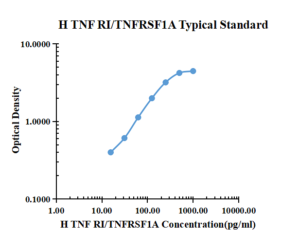
| pg/ml | O.D. | Average | Corrected | |
|---|---|---|---|---|
| 0.00 | 0.0711 | 0.0713 | 0.0712 | |
| 15.63 | 0.5380 | 0.4053 | 0.4717 | 0.4005 |
| 31.25 | 0.6768 | 0.6877 | 0.6823 | 0.6111 |
| 62.50 | 1.1630 | 1.2380 | 1.2005 | 1.1293 |
| 125.00 | 2.0080 | 2.1190 | 2.0635 | 1.9923 |
| 250.00 | 3.1820 | 3.3390 | 3.2605 | 3.1893 |
| 500.00 | 4.3384 | 4.2418 | 4.2901 | 4.2189 |
| 1000.00 | 4.4053 | 4.6583 | 4.5318 | 4.4606 |
Product Features
- Optimized capture and detection antibody pairings with recommended concentrations save lengthy development time
- Development protocols are provided to guide further assay optimization
- Assay can be customized to your specific needs
- Economical alternative to complete kits
Kit Content
- Capture Antibody
- Detection Antibody
- Recombinant Standard
- Streptavidin conjugated to horseradish-peroxidase (Streptavidin-HRP)
Other Reagents Required
DTSet Ancillary Reagent Kit (5 plates): containing 96 well microplates, plate sealers, substrate solution, stop solution, plate coating buffer (PBS), wash buffer, and assay buffer.
- 96 well microplates: YOUKE Life, Catalog # DSEP01. Plate Sealers: YOUKE Life, Catalog # DSSF01.
- Coating Buffer: 137 mM NaCl, 2.7 mM KCl, 8.1 mM Na2HPO4, 1.5 mM KH2PO4, pH 7.2-7.4, 0.2μm filtered . YOUKE Life, Catalog # DSCB01.
- Blocking Buffer: YOUKE Life, Catalog # DSBB01.
- Wash Buffer: 0.05% Tween® 20 in PBS, pH 7.2-7.4. YOUKE Life, Catalog # DSWB01.
- Assay Buffer: 0.5% BSA,0.05% Tween® 20,PBS Solution.YOUKE Life, Catalog # DSAB01
- Substrate Solution: Tetramethylbenzidine. YOUKE Life, Catalog # DSTS01.
- Stop Solution: 0.5mol/ml H2SO4. YOUKE Life, Catalog # DSSS01.
Product Data Sheet
Background: TNF RI
Tumor necrosis factors (TNFs) are pleiotropic cytokines that are considered primary modifiers of the inflammatory and immune reactions of animals produced in response to injury or infection. Two forms of TNF, designated TNF-alpha (or cachectin) and TNF-beta (or lymphotoxin), have been described that share 30% sequence similarity and compete for binding to the same receptors. TNFs play a necessary and beneficial role as mediators of host resistance to infections and tumor formation. However, over-production or inappropriate expression of these factors can lead to a variety of pathological conditions, including wasting, systemic toxicity, and septic shock.
The actions of TNFs are produced subsequent to binding of the factors to cell surface receptors. Two distinct TNF receptors have been identified and cloned. Virtually all cell types studied show the presence of one or both of these receptor types. One receptor type, termed TNF RII (Type A, Type a, 75 kDa or utr antigen), shows an apparent molecular weight of 75 kDa. The gene for this receptor encodes a presumptive transmembrane protein of 439 amino acid (aa) residues. The other receptor type, termed TNF RI (Type B, Type b, 55 kDa or htr antigen), shows an apparent molecular weight of 55 kDa. The gene for this protein encodes a transmembrane protein of 426 aa residues. Both receptor types show high affinity binding of either TNF-alpha or TNF-beta. The two receptor types are immunologically distinct but their extracellular domains show similarities in the pattern of cysteine residue locations in four domains. The intracellular domains of the two receptor types are apparently unrelated, suggesting the possibility that the two receptor types employ different signal transduction pathways.
Several groups have identified soluble TNF binding proteins in human serum and urine that can neutralize the biological activities of TNF-alpha and TNF-beta. Two types have been identified and designated sTNF RI (or TNF BPI) and sTNF RII (or TNF BPII). These soluble forms have now been shown to represent truncated forms of the two types of TNF receptors discussed above. The soluble receptor forms apparently arise as a result of shedding of the extracellular domains of the receptors, and concentrations of about 1-2 ng/mL are found in the serum and urine of healthy subjects. The levels of the soluble receptors vary from individual to individual but are stable over time for given individuals.
Elevated levels of TNF receptors have been found in the amniotic fluid and urine of pregnant women, in serum or plasma in association with pathological conditions such as endotoxinemia, meningiococcemia, and HIV infection, and in plasma and ascites of patients in association with infections and malignancies. The mechanisms involved in the induction of shedding of the TNF receptors are not well understood. There are reports of correlations between increased TNF levels and soluble receptor levels, suggesting generally that stimuli that cause TNF levels to rise also induce shedding of TNF receptors. There is also evidence, however, that suggests the shedding of the two types of soluble receptors is independently regulated. 2 For research use only. Not for use in diagnostic procedures.
The physiological role of the soluble TNF receptors is not known. It is known that both types of soluble receptors can bind to TNF in vitro and inhibit its biological activity by competing with cell surface receptors for TNF binding. Consequently it has been suggested that shedding of soluble receptors in response to TNF release could serve as a mechanism for binding and inhibiting the TNF not immediately bound to surface receptors, thus protecting other cells from the effects of TNF and localizing the inflammatory response. It is also possible that shedding of receptors represents a mechanism for desensitizing the cells that shed the receptors from the effects of TNF. On the other hand, it has been reported that at low concentrations of TNF, binding to soluble receptors can stabilize TNF and augment some of its activities. Thus it is possible that under some conditions the pool of TNF bound to soluble receptors could represent a reservoir for the stabilization and controlled release of TNF.
