Human M-CSF enzyme-linked immunoassay kit
| Specification | 96 Test |
|---|---|
| Sensitivity | 1.41 pg/ml (50 μl);15.89 pg/ml (10 μl) |
| Standard Curve Range | 78.13~5000 pg/ml |
| Standard Curve Gradient | 7 Points |
| Number of Incubations | 2 |
| Detectable sample | Liquid phase sample of soluble substances. For example: serum, plasma, cell culture supernatant, tissue grinding liquid, etc. |
| Sample Volume | 50 μl/10 μl |
| Type | Fully Ready-to-Use |
| Operation Duration | 120min |
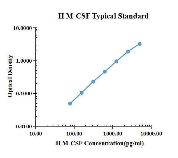
| pg/ml | O.D. | Average | Corrected | |
|---|---|---|---|---|
| 0.00 | 0.0157 | 0.0157 | 0.0157 | |
| 78.13 | 0.0691 | 0.0610 | 0.0651 | 0.0494 |
| 156.25 | 0.1317 | 0.1102 | 0.1210 | 0.1053 |
| 312.50 | 0.2432 | 0.2484 | 0.2458 | 0.2301 |
| 625.00 | 0.4781 | 0.4755 | 0.4768 | 0.4611 |
| 1250.00 | 0.9490 | 0.9950 | 0.9720 | 0.9563 |
| 2500.00 | 2.0360 | 1.7810 | 1.9085 | 1.8928 |
| 5000.00 | 3.3130 | 3.2000 | 3.2565 | 3.2408 |
Precision
| Intra-assay Precision | Inter-assay Precision | |||||
| Sample Number | S1 | S2 | S3 | S1 | S2 | S3 |
| 22 | 22 | 22 | 6 | 6 | 6 | |
| Average(pg/ml) | 165.6 | 689.8 | 2146.1 | 110.7 | 548.3 | 1660.5 |
| Standard Deviation | 9.8 | 42.2 | 119.7 | 5.2 | 37.6 | 83.2 |
| Coefficient of Variation(%) | 5.9 | 6.1 | 5.6 | 4.7 | 6.9 | 5.0 |
Intra-assay Precision (Precision within an assay) Three samples of known concentration were tested Twenty-two times on one plate to assess intra-assay precision.
Inter-assay Precision (Precision between assays) Three samples of known concentration were tested six times on one plate to assess intra-assay precision.
Spike Recovery
The spike recovery was evaluated by spiking 3 levels of human M-CSF into health human serum sample. The un-spiked serum was used as blank in this experiment.
The recovery ranged from 72% to 121% with an overall mean recovery of 105%.
Sample Values
| Sample Matrix | Sample Evaluated | Range (pg/ml) | Detectable (%) | Mean of Detectable (pg/ml) |
|---|---|---|---|---|
| Serum | 30 | 684.79-1505.39 | 100 | 1024.43 |
Serum/Plasma – Thirty samples from apparently healthy volunteers were evaluated for the presence of CD137/4-1BB/TNFRSF9 in this assay. No medical histories were available for the donors.
Product Data Sheet
Background: M-CSF
M-CSF, also known as CSF-1, is a four-alpha -helical-bundle cytokine that is the primary regulator of macrophage survival, proliferation and differentiation. M-CSF protein is also essential for the survival and proliferation of osteoclast progenitors. M-CSF also primes and enhances macrophage killing of tumor cells and microorganisms, regulates the release of cytokines and other inflammatory modulators from macrophages, and stimulates pinocytosis. M-CSF increases during pregnancy to support implantation and growth of the decidua and placenta. Sources of M-CSF include fibroblasts, activated macrophages, endometrial secretory epithelium, bone marrow stromal cells and activated endothelial cells . The M-CSF receptor (c-fms) transduces its pleotropic effects and mediates its endocytosis. M-CSF mRNAs of various sizes occur. Full length mouse M-CSF transcripts encode a 520 amino acid (aa) type I transmembrane (TM) protein with a 462 aa extracellular region, a 21 aa TM domain, and a 37 aa cytoplasmic tail that forms a 140 kDa covalent dimer. Differential processing produces two proteolytically cleaved, secreted dimers. One is an N- and O- glycosylated 86 kDa dimer, while the other is modified by both glycosylation and chondroitin-sulfate proteoglycan (PG) to generate a 200 kDa subunit. Although PG-modified M-CSF protein can circulate, it may be immobilized by attachment to type V collagen . Shorter transcripts encode M‑CSF that lacks cleavage and PG sites and produces an N-glycosylated 68 kDa TM dimer and a slowly produced 44 kDa secreted dimer. Although forms may vary in activity and half-life, all contain the N-terminal 150 aa portion that is necessary and sufficient for interaction with the M-CSF receptor. The first 229 aa of mature mouse M-CSF shares 87%, 83%, 82% and 81% aa identity with corresponding regions of rat, dog, cow and human M-CSF, respectively. Human M‑CSF is active in the mouse, but mouse M-CSF is reported to be species-specific.
