Human M-CSF DTSet enzyme-linked immunoassay kit
| Specification | 96*5 Test;96T*15 Test |
|---|---|
| Standard Curve Range | 15.63 pg/ml -1000 pg/ml |
| Standard Curve Gradient | 7 Points/3 Folds |
| Number of Incubations | 2 |
| Detectable sample | Liquid phase sample of soluble substances. For example: serum, plasma, cell culture supernatant, tissue grinding liquid, etc. |
| Sample Volume | 50 μl |
| Type | Not Ready-to-Use |
| Test Duration | 120min |
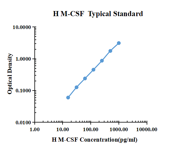
| pg/ml | O.D. | Average | Corrected | |
|---|---|---|---|---|
| 0.00 | 0.0474 | 0.0458 | 0.0466 | |
| 15.63 | 0.1091 | 0.1044 | 0.1068 | 0.0602 |
| 31.25 | 0.1705 | 0.1756 | 0.1731 | 0.1265 |
| 62.50 | 0.2885 | 0.2840 | 0.2863 | 0.2397 |
| 125.00 | 0.4956 | 0.5032 | 0.4994 | 0.4528 |
| 250.00 | 0.9218 | 0.9077 | 0.9148 | 0.8682 |
| 500.00 | 1.8180 | 1.7930 | 1.8055 | 1.7589 |
| 1000.00 | 3.1670 | 3.1440 | 3.1555 | 3.1089 |
Product Features
- Optimized capture and detection antibody pairings with recommended concentrations save lengthy development time
- Development protocols are provided to guide further assay optimization
- Assay can be customized to your specific needs
- Economical alternative to complete kits
Kit Content
- Capture Antibody
- Detection Antibody
- Recombinant Standard
- Streptavidin conjugated to horseradish-peroxidase (Streptavidin-HRP)
Other Reagents Required
DTSet Ancillary Reagent Kit (5 plates): containing 96 well microplates, plate sealers, substrate solution, stop solution, plate coating buffer (PBS), wash buffer, and assay buffer.
- 96 well microplates: YOUKE Life, Catalog # DSEP01. Plate Sealers: YOUKE Life, Catalog # DSSF01.
- Coating Buffer: 137 mM NaCl, 2.7 mM KCl, 8.1 mM Na2HPO4, 1.5 mM KH2PO4, pH 7.2-7.4, 0.2μm filtered . YOUKE Life, Catalog # DSCB01.
- Blocking Buffer: YOUKE Life, Catalog # DSBB01.
- Wash Buffer: 0.05% Tween® 20 in PBS, pH 7.2-7.4. YOUKE Life, Catalog # DSWB01.
- Assay Buffer: 0.5% BSA,0.05% Tween® 20,PBS Solution.YOUKE Life, Catalog # DSAB01
- Substrate Solution: Tetramethylbenzidine. YOUKE Life, Catalog # DSTS01.
- Stop Solution: 0.5mol/ml H2SO4. YOUKE Life, Catalog # DSSS01.
Product Data Sheet
Background: M-CSF
M-CSF, also known as CSF-1, is a four-alpha -helical-bundle cytokine that is the primary regulator of macrophage survival, proliferation and differentiation. M-CSF protein is also essential for the survival and proliferation of osteoclast progenitors. M-CSF also primes and enhances macrophage killing of tumor cells and microorganisms, regulates the release of cytokines and other inflammatory modulators from macrophages, and stimulates pinocytosis. M-CSF increases during pregnancy to support implantation and growth of the decidua and placenta. Sources of M-CSF include fibroblasts, activated macrophages, endometrial secretory epithelium, bone marrow stromal cells and activated endothelial cells . The M-CSF receptor (c-fms) transduces its pleotropic effects and mediates its endocytosis. M-CSF mRNAs of various sizes occur. Full length mouse M-CSF transcripts encode a 520 amino acid (aa) type I transmembrane (TM) protein with a 462 aa extracellular region, a 21 aa TM domain, and a 37 aa cytoplasmic tail that forms a 140 kDa covalent dimer. Differential processing produces two proteolytically cleaved, secreted dimers. One is an N- and O- glycosylated 86 kDa dimer, while the other is modified by both glycosylation and chondroitin-sulfate proteoglycan (PG) to generate a 200 kDa subunit. Although PG-modified M-CSF protein can circulate, it may be immobilized by attachment to type V collagen . Shorter transcripts encode M‑CSF that lacks cleavage and PG sites and produces an N-glycosylated 68 kDa TM dimer and a slowly produced 44 kDa secreted dimer. Although forms may vary in activity and half-life, all contain the N-terminal 150 aa portion that is necessary and sufficient for interaction with the M-CSF receptor. The first 229 aa of mature mouse M-CSF shares 87%, 83%, 82% and 81% aa identity with corresponding regions of rat, dog, cow and human M-CSF, respectively. Human M‑CSF is active in the mouse, but mouse M-CSF is reported to be species-specific.
